
Understanding molecular mechanisms of neuroprotection
The goal of retinal neuroprotection is to preserve the structure and function of neurons, preventing or slowing the progression of vision loss by maintaining the health of retinal neurons and their connections. There is an ongoing quest for neuroprotective factors that can prevent or slow down the progression of retinal degeneration.
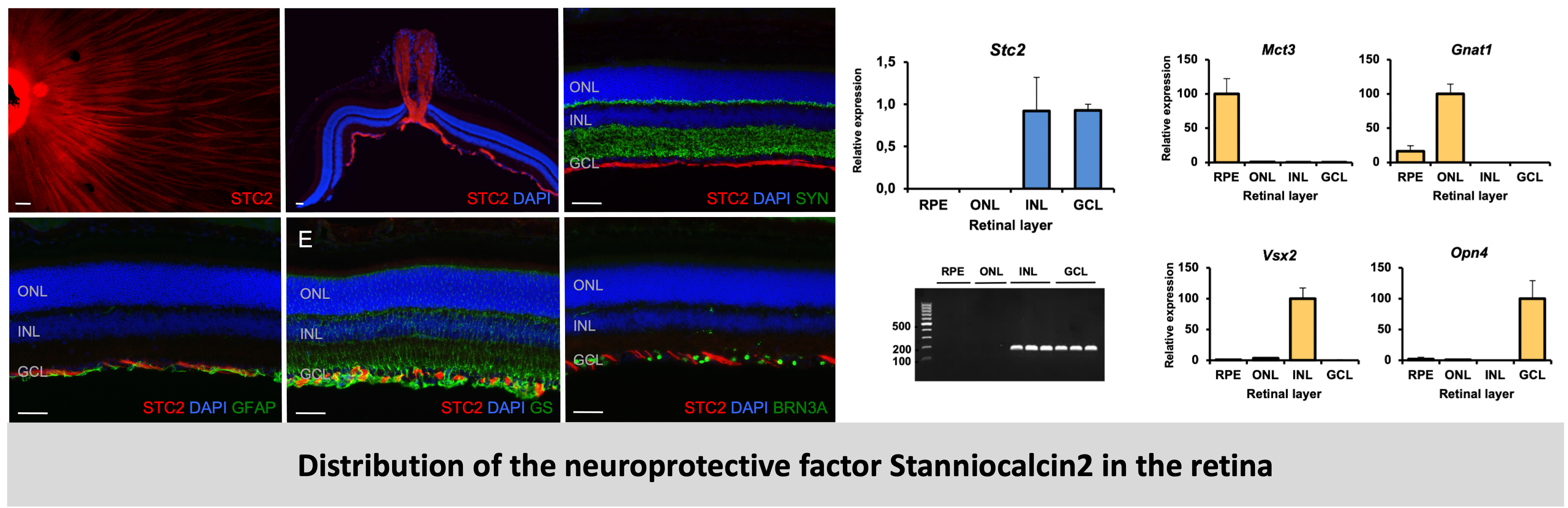
Hypoxia-mediated neuroprotection: Chronic (long-term) hypoxia causes retinal degeneration, however acute (short-term) hypoxic stress has been shown to provide transient protection against subsequent damage in the retina. Stanniocalcins – STC1 and STC2 – are secreted glycoproteins that are hypoxia-regulated and were shown to be cytoprotective in various in vitro studies. Hence, we investigated the expression of stanniocalcins in the normal, degenerating and hypoxic retina. Using wild-type and Rd10 (Retinal degeneration model) mice, we showed that both Stc2 and its paralog Stc1 are expressed in the developing and the aging retina, but only Stc2 is hypoxia responsive and regulated in a HIF1A- dependent manner. Retinal expression of Stc2, but not Stc1, was induced in mice in an in vivo model of acute hypoxia and a genetic model of chronic hypoxia. Using laser capture microdissection and immunofluorescence, we showed that surprisingly, Stc2 was not normally expressed in photoreceptors but in the inner retina, specifically in the inner nuclear layer (INL) and the ganglion cell layer (GCL), and the protein localized to the neurofilaments. The expression of both Stc1 and Stc2 remained unchanged in the degenerative retina that had an almost complete loss of photoreceptors, confirming their expression in the inner retina. In collaboration with a team from the University of Sydney, Australia that had developed stanniocalcin knock-out mice (Stc2-/- and Stc1-/-), we analyzed the retinal morphologies of both the Stc1 and Stc2 knockout mice. The absence of either Stc1 or Stc2 had no effect on retinal architecture, as was evident from retinal morphology of the respective knockout mice indicating that Stc2 may not be essential for the normal development of the retina, but only involved in hypoxic stress mechanisms. This study provided evidence for the differential hypoxia-mediated regulation of STC1 and STC2 in the retina. STC2 may play a role in the retina's response to hypoxic stress contributing to neuroprotection during retinal degeneration and hence has strong potential as a candidate retinal neuroprotective factor.
Read the details and the results of this project here
Stanniocalcin2, but not Stanniocalcin1, responds to hypoxia in a HIF1-dependent manner in the retina
Ail D, Samardzija M, Chang ACM, Keck J, Reddel RR., Grimm C
Frontiers in Neuroscience, 2022
Glia-mediated neuroprotection: The retina is a complex tissue composed of many cell types. In addition to the neurons that are present in three distinct stratified layers, the retina is also composed of epithelial cells present as the retinal pigment epithelium (RPE) and glial cells (Muller cells, astrocytes and microglia). Glial cells of the retina provide structural, metabolic, and functional support to retinal neurons. They help maintain homeostasis, regulate synaptic activity, and protect the retina from damage. In many blinding diseases of the retina, loss of function and thus severe visual impairment results from apoptotic cell death of damaged photoreceptors. In an attempt to survive, injured photoreceptors generate survival signals to induce intercellular protective mechanisms that eventually may rescue photoreceptors from entering an apoptotic death pathway. One such endogenous survival pathway is controlled by leukemia inhibitory factor (LIF), which is produced by a subset of Muller glia cells in response to photoreceptor injury. In the absence of LIF, survival components are not activated and photoreceptor degeneration is accelerated. Although LIF is a crucial factor for photoreceptor survival, the detailed mechanism of its induction in the retina has not been elucidated. Through this study, we showed that administration of tumor necrosis factor-alpha (TNF) was sufficient to fully upregulate Lif expression in Muller cells in vitro and in the retina in vivo. Increased Lif expression depended on p38 mitogen-activated protein kinase (MAPK), since inhibition of its activity suppressed Lif expression in vitro and in vivo. Inhibition of p38 MAPK activity reduced the Lif expression also in the model of light-induced retinal degeneration and resulted in increased cell death in the light-exposed retina. Thus, expression of Lif in the injured retina and activation of the endogenous survival pathway involve signaling through p38 MAPK. Since p38 MAPK positively regulates LIF-mediated neuroprotection, modulating this pathway could be a strategy to slow retinal degeneration.
Read the details and the results of this project here
P38 MAPK signaling acts upstream of LIF-dependent neuroprotection during photoreceptor degeneration.
Agca C, Gubler A, Traber G, Beck C, Imsand C, Ail D, Caprara C, Grimm C
Cell Death and Disease, 2013