
Understanding molecular mechanisms of regeneration
The retina is a light-sensitive tissue at the back of the eye that plays a critical role in visual signal processing. Retinal regeneration is the process by which damaged or lost retinal cells—such as photoreceptors, or other neurons—are replaced or repaired. Some species of fish (eg. Zebrafish) and frogs (eg. Xenopus laevis) can regenerate their retina, which means if these species lose their photoreceptors, they can grow them back and restore vision. However, in most mammals, including humans, the retina has very limited regenerative capacity, making degenerative diseases like retinitis pigmentosa, age-related macular degeneration (AMD), and glaucoma irreversible. Understanding why frogs can regenerate their retina while humans don’t could lead to development of new regenerative therapies for humans.

Read more about mechanisms of regeneration here
Retinal degeneration and regeneration – Lessons from fishes and amphibians
Ail D and Perron M
Current Pathobiology Reports, 2017
Muller glia cell-mediated regeneration
The retina is a complex tissue composed of many cell types. In addition to the neurons that are present in three distinct stratified layers, the retina is also composed of epithelial cells present as the retinal pigment epithelium (RPE) and glial cells (Muller cells, astrocytes and microglia). Müller glial cells are the principal support cells of the retina, providing structural, metabolic, and neuroprotective functions.
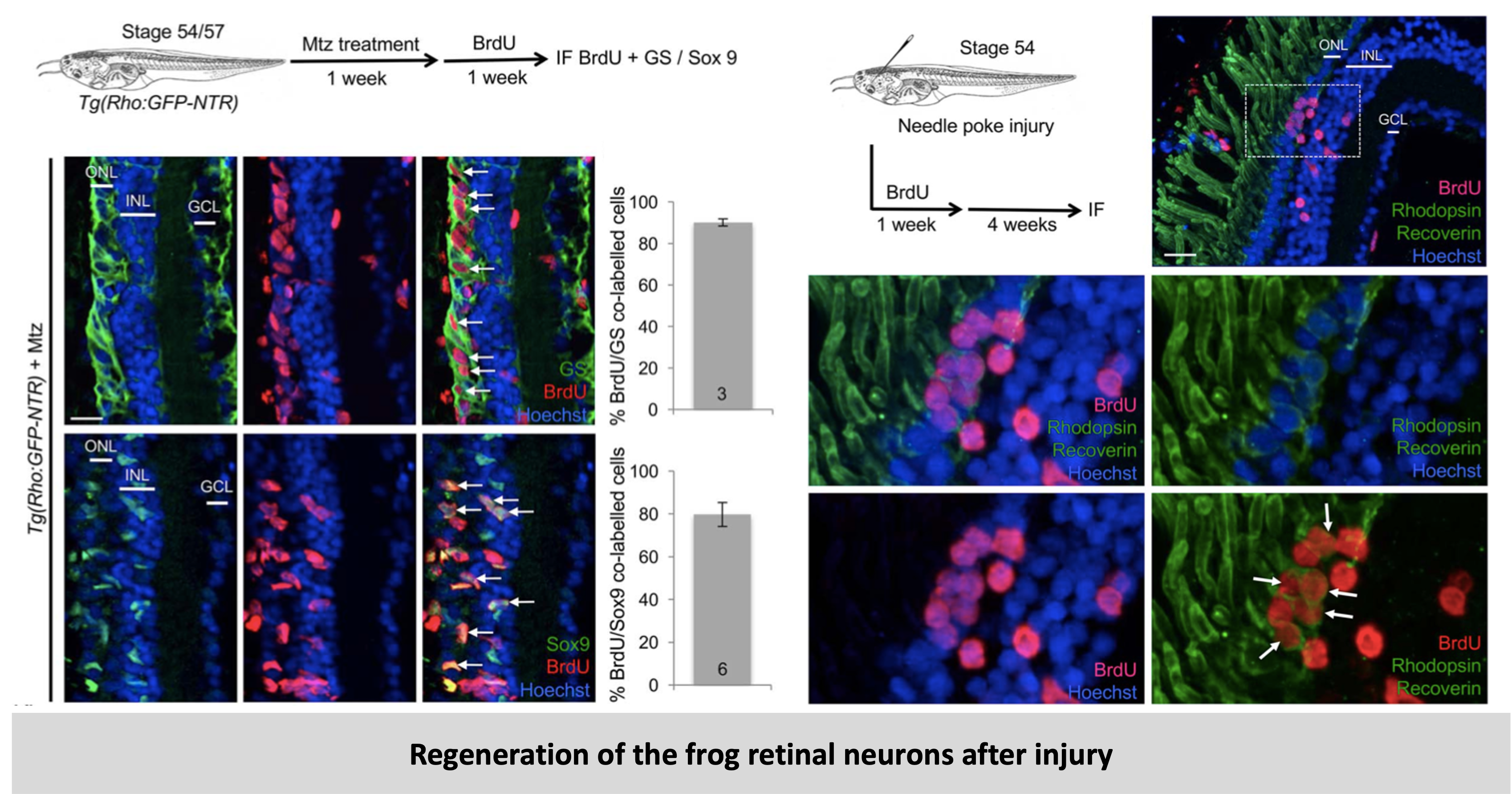
If the frog species Xenopus laevis loses its retinal cells due to damage or degeneration, it can grow them back (meaning the frog can regenerate its retina). It is capable of doing so because of a pool of stem cells present in a special zone called CMZ (Ciliary Marginal Zone). A CMZ-like structure or a stem cell pool is not present in human retina. However, other cell types in the frog retina (RPE and Muller cells which are also present in humans) have shown some regenerative potential. As the regenerative potential of Muller glia cells was unexplored in Xenopus, we tackled this issue using two Xenopus retinal injury paradigms: a physical injury model (local damage model) and a transgenic model allowing for conditional photoreceptor cell ablation (global degeneration model). Using these models, we were able to show that when the photoreceptors are damaged or degenerated in the frog species – Xenopus laevis, Müller cells play a key role in retinal regeneration by reprogramming into retinal progenitor-like cells that can divide and replace the damaged neurons.
Read the details and the results of this project here
Müller glial cell reactivation in Xenopus models of retinal degeneration.
Langhe R*, Chesneau A*, Colozza G, Hidalgo M, Ail D, Locker M, Perron M
Glia, 2016
Identification of a novel factor responsible for regeneration
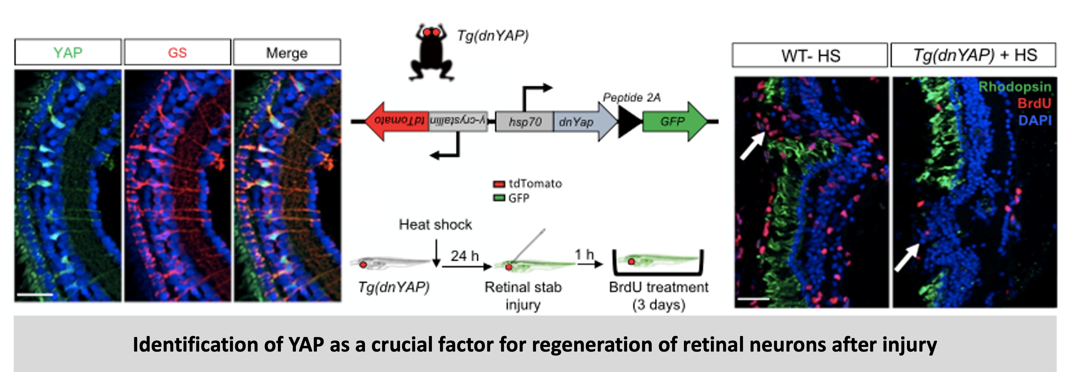
Normally following damage or degeneration the Xenopus retina regenerates using the endogenous stem cell pool. However, we showed that Müller glia cells also proliferate and regenerate photoreceptors in Xenopus. To further understand the mechanisms of regeneration and the factors involved we examined the Hippo/YAP pathway, which recently emerged as a crucial player in the field of stem cell biology and regeneration. We found that the key transcription factor of this pathway called YAP is specifically expressed in Müller cells. We further examined the involvement of YAP by taking advantage of Xenopus transgenic lines for inducible overexpression or knock down of YAP. We showed that knock-down of YAP in the Muller cells suppressed the regeneration potential of the Xenopus retina. YAP activity stimulation can enhance Müller cell proliferation and retinal repair in injured or degenerated retina. This study highlights a YAP-EGFR (epidermal growth factor receptor) axis by which Müller cells exit their quiescent state, a critical step toward regeneration. In the mouse retina, where Müller cells do not spontaneously proliferate, overactivation of YAP is sufficient to induce their reprogramming into highly proliferative cells. These findings suggest that modulating YAP activity could be a potential therapeutic strategy for promoting retinal regeneration in degenerative diseases.
Read the details and the results of this project here
Linking YAP to Muller glia quiescence exit in the degenerative retina.
Hamon A*, Ail D*, Garcia D*, Bitard J, Chesneau A, Dalkara D, Locker M, Roger J, Perron M
Cell Reports, 2019
The projects on retinal regeneration were conducted in the ScaNR Lab (Stem Cell and Neurogenesis in the Retina) which is part of the Paris-Saclay Institute of Neuroscience (NeuroPSI) at the University of Paris-Saclay. The lab is mainly involved in the study of retinal regeneration and my focus was on elucidating the role of YAP in Muller-cell mediated regeneration of the Xenopus retina.